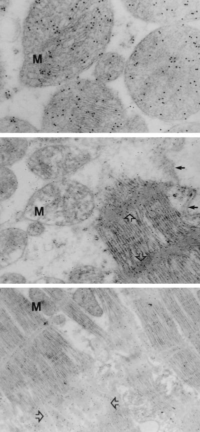
Figure 2.
Ultrastructural localization of cytochrome c. High magnifications of normal myocardial specimen from a donor heart (Top) and ischemic cardiomyopathic hearts (Middle and Bottom) demonstrate striking difference in cytochrome c immunoreactivity. Localization of cytochrome c is represented by (black) gold particles. The cytochrome c is predominantly localized in mitochondria (M) in normal hearts (Top). On the other hand, cytochrome c is substantially reduced in mitochondria (Middle, M) in cardiomyopathic heart and is concentrated either over Z-bands (Middle, open arrows) or near intercalated disc (Bottom, open arrows). Small arrows indicate the presence of normal sarcolemma (Middle).

