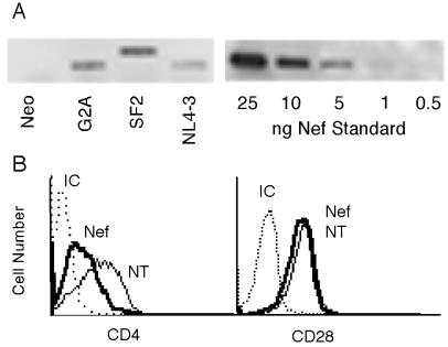
Figure 1.
Physical and functional confirmation of Nef expression. Western blot analysis (A) shows Nef expression from Jurkat E6–1 T cells. The HIV-1 nef gene was introduced into cells by liposome-mediated LXSN retroviral transduction, followed by selection in G418. Cells were transduced with nonmyristylated NL4–3 Nef (G2A), myristylated NL4–3 Nef (NL4–3), and SF2 Nef (SF2) and by the LXSN control vector (Neo). Cells were lysed and Nef was immunoprecipitated with rabbit anti-Nef polyclonal antibody. Samples from 5 million cells were electrophoresed, and resolved proteins were transferred to nitrocellulose. Nef was detected by monoclonal anti-Nef antibody. A Western blot for recombinant NL4–3 Nef is shown at 0.5–25 ng per lane. (B) FITC analysis of cell surface expression of CD28 and CD4 on nontransduced (NT) cells (thin line) or on cells transduced with HIV-1 Nef from strain SF2 (thick line). Cells were stained directly with FITC-conjugated anti-human mAbs or with murine isotype control Abs (IC).

