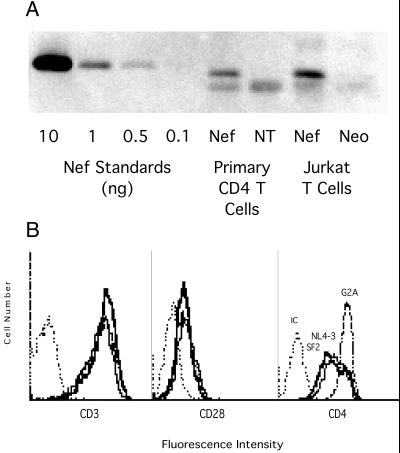
Figure 4.
Physical and functional confirmation of Nef expression in primary CD4 T cells. Western blot analysis (A) compares NL4–3 Nef expression in transduced primary CD4 T cells and Jurkat cells, in addition to control non-Nef (NT or Neo) cells. The blot also includes recombinant NL4–3 Nef at 0.1–10 ng/lane. (B) Flow cytometric analysis of cell surface expression of CD3, CD28, and CD4 on cells transduced with HIV-1 Nef from strain SF2, NL4–3, or the nonmyristylated NL4–3 (G2A) as shown. Cells were stained directly with FITC-conjugated anti-human mAbs or with murine isotype control Abs (IC). Viability (dye exclusion) of SF2, NL4–3, G2A, and NT cells was 88, 91, 92, and 83%, respectively. Proliferation (doubling time) of cell populations was 2.9, 2.6, 3.5, and 2.6 days, respectively.

