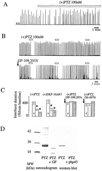
Figure 2.
Desensitization of the σ1 response via cPKC. (A) Desensitization of the σ1 effect on the integrated XII activity after 30 min of perfusion (open bar). (B) Desensitization induced by successive, short perfusions of PTZ (Upper) was prevented by a preperfusion of GF-109,203x (50 nM, 5 min; Lower). (C) Mean values of the effects of successive, short perfusions of σ1 in control conditions and after GF-109,203x and Gö-6976 perfusions, which prevented the PTZ-induced desensitization. (D) Autoradiogram after a long perfusion of PTZ revealed a phosphorylated, 28-kDa protein at the level of the σ1 receptor protein (revealed by Western blot analysis), which did not appear after GF-109,203x perfusion. In the Western blot, this band disappeared in the presence of the synthetic peptide anti-PBP45 (pbp-45) (3).

