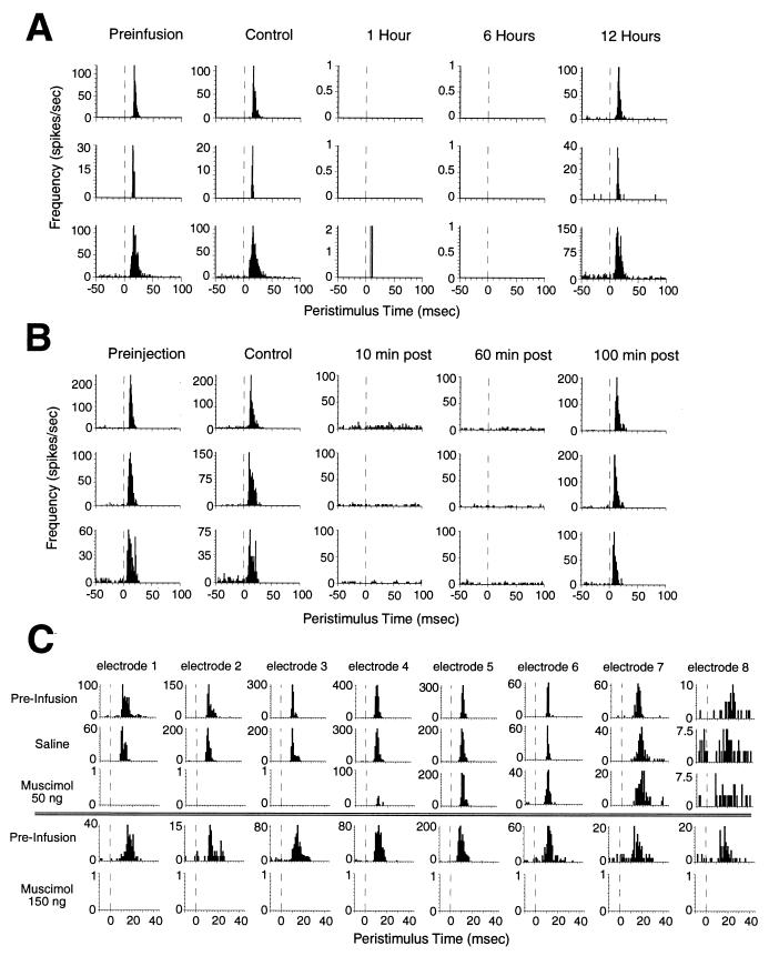
Figure 2.
(A) Examples of the duration and reversibility of muscimol inactivation. Peristimulus time histograms (PSTHs) for three SI neurons resulting from repetitively stimulating a single whisker before (preinfusion), after infusion of saline vehicle (150 nl) into the SI cortex (control), and 1, 6, and 12 h after infusion of muscimol (150 ng) into the SI cortex. Although infusion of saline vehicle had no effect on cortical activity, muscimol completely abolished cortical activity for approximately 9 h. The inactivation was completely reversible: responses returned to baseline within 12 h. (B) Examples of the duration and reversibility of lidocaine-induced peripheral deafferentation. PSTHs for three VPM neurons before any lidocaine injections (preinjection), after a control injection, and 10, 60, and 100 min after a subcutaneous injection (20 μl, 1%) near the stimulated whisker. The effective lidocaine injection completely anesthetized the stimulated region of the face for at least 60 min, and the effect was completely reversible. (C) Spatial extent of muscimol inactivation of the SI cortex. Each of the eight columns represents the PSTH from a single neuron isolated on each of the eight microwires of an SI cortical array after stimulation of a single whisker. The muscimol infusion cannula is 250 μm away from microelectrode 1 (microwires extended linearly away from the cannula, each wire 250 μm away from the adjacent wire, see Fig. 1A). Infusion of saline had no effect on neural activity on any of the wires. A low dose of muscimol (50 ng in 50 nl of saline) abolished activity on three or four wires (approximately 1.2 mm). In a different experiment (Lower), a higher dose of muscimol (150 ng in 150 nl of saline) abolished neural activity on all microwires (approximately 2.2 mm). This dose (150 ng) effectively abolished activity throughout the barrel region of the SI cortex and was used in all experiments involving facial lidocaine. The functional spread of each muscimol infusion during each experiment was confirmed by monitoring activity on the cortical array.

