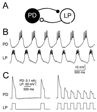
Figure 1.
The PD and LP neurons of the pyloric network make reciprocally inhibitory synapses. (A) Schematic diagram of PD and LP neurons and their connectivity. (B) Simultaneous intracellular recordings of LP and PD in normal saline shows alternating activity. (C) Voltage step depolarizations of the LP neuron in tetrodotoxin produces a graded IPSC in the PD neuron. During each voltage pulse, the IPSC depresses; it recovers between pulses. The recovery from depression is a direct function of the time interval between pulses. The LP neuron was voltage clamped with a holding potential of −60 mV. The PD neuron was voltage clamped at a constant potential of −35 mV and the synaptic currents were measured. B and C are recordings from the same experimental preparation.

