
Figure 3.
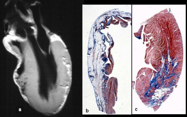
Same case of fig. 2. Note the biventricular involvement at long axis in vitro MRI (a), with transmural fibro-fatty replacement in the RV free wall (b) and focal subepicardial in the LV free wall (c).
Official websites use .gov
A
.gov website belongs to an official
government organization in the United States.
Secure .gov websites use HTTPS
A lock (
) or https:// means you've safely
connected to the .gov website. Share sensitive
information only on official, secure websites.
Same case of fig. 2. Note the biventricular involvement at long axis in vitro MRI (a), with transmural fibro-fatty replacement in the RV free wall (b) and focal subepicardial in the LV free wall (c).