Figure 1.
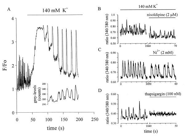
(A) Effect of high K+ on [Ca2+]i oscillations and contractions observed with CLSM in a single stage-2 cardiomyocyte. Depolarization resulted in a transient rise of [Ca2+]i, presumably because of the activation of VDCC channels and in an alteration of the amplitude and frequency of oscillations. The inset shows cell contractions time-locked with [Ca2+]i oscillations during superfusion with high K+. Cell membrane depolarization resulted in an initial discontinuation of contractions, which resumed with the decline of [Ca2+]i. F/Fo indicates the ratio between the actual and the initial intensity of the fluorescent dye. Grey-levels represent the intensity of the transmitted light, which varied with cell contraction. (B–D) Effect of different pharmacological agents on 340/380 ratios in fura-2AM-loaded spontaneously beating stage-2 cells. (B) The VDCC blocker nisoldipine (2 μM) did not impair [Ca2+]i oscillations but lead to a lowering of steady-state [Ca2+]i levels. (C) Similarly, Ni2+ (2 mM), known to block the Na+-Ca2+ exchanger, did not interrupt [Ca2+]i oscillations. (D) The Ca2+-ATPase inhibitor thapsigargin (100 nM) led to an abrupt halt of the [Ca2+]i oscillations and a concomitant increase of the resting [Ca2+]i.

