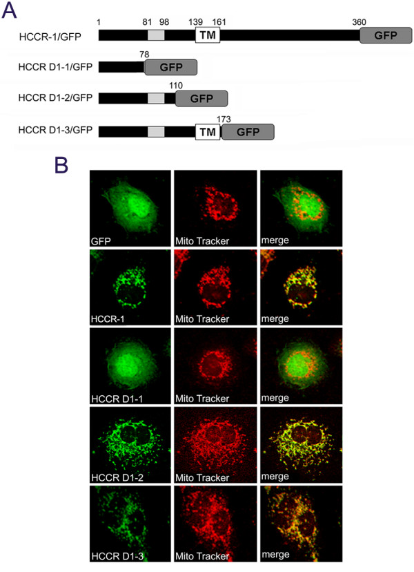
Figure 3.

NH2-terminus of HCCR-1 contains the mitochondrial targeting information. (A) Schematic structure of HCCR-1 and its deleted mutants which were COOH-terminally linked by the GFP reporter gene. Grey boxes indicate the hydrophobic region. TM boxes indicate the locations of putative transmembrane domain. (B) Expression of wild-type or deleted mutants in COS-7 cells. COS-7 cells were transfected with the indicated constructs in the expression vectors. The cells were incubated with MitoTracker and fixed. Fluorescent images of GFP (green), MitoTracker (red) were taken using a confocal microscope. Merged fluorescent images of GFP and MitoTracker are shown. Other conditions are described in Materials and methods.
