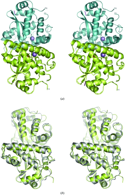
Figure 2.
(a) Stereoview of a ribbon representation of the structure of a physiologically relevant Mn-SODDr dimer. The two monomers making up the dimer are coloured lime-green (with Cα also shown) and cyan, respectively, with the positions of the Mg3+ ions shown in magenta. The C-terminus of each monomer is coloured red and the N-terminus is blue. Residues forming part of the six-residue QGQNGA insert are numbered to show the likely position of disordered residues with respect to the rest of the protein. (b) A superposition of the CD dimer found in crystal form II of Mn-SODDr and the dimer found in the crystal structure of Mn-SODEc. This shows that both monomers and dimers of the two Mn-SODs are essentially isostructural. The figure was produced using PyMOL (DeLano, 2002 ▶).

