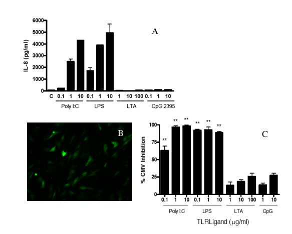
Figure 1.

IL-8 secretion and HCMV inhibition in HFF induced by TLR ligands. HFF cells were cultured to 95% confluency and stimulated with the indicated doses of TLR ligands or medium control alone (C) for 24 hours. A. Culture supernatants were then collected and assayed for IL-8 by ELISA. IL-8 secretion from one experiment representative of three. Bars represent mean ± SD of triplicate cultures. B and C. After treatment of HFF with medium alone, LTA, Poly I:C, LPS, or CpG 2395 for 24 hours, cells were washed and CMVPT30-gfp was added. After four hours, the virus innoculum was removed and replaced with fresh culture medium. HCMV infection was quantified on day 10 post-infection by counting fluorescent (GFP expressing) cells in each well. B. Shown is a representative culture well from cells treated with medium alone. C. Percent inhibition compared to medium control. Results of one experiment, representative of 3 independent experiments, is shown. Bars represent mean ± SD of triplicate cultures. * indicates P ≤ 0.05 compared to control. ** indicates P ≤ 0.01 compared to control. *** indicates P ≤ 0.001 compared to control.
