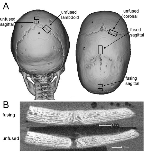
Figure 1.

Computer tomography (CT) scans showing site and fusion state of sutures obtained from craniosynostosis patients. A) Posterior and superior (left and right) view of patient #58 indicating where unfused, fusing and fused sutures were obtained from. p, parietal bone; o, occipital bone; f, frontal bone. B) MicroCT image demonstrating a fusing and unfused suture. Scale 1 mm.
