
Figure 8.
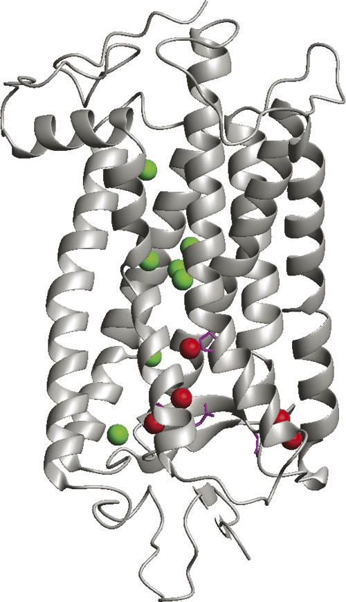
Structure of bovine rhodopsin showing positions of mutations associated with autosomal dominant retinitis pigmentosa (ADRP). Red spheres mark positions of five buried waters which contact sites of ADRP mutations thought to cause rhodopsin misfolding. Green spheres show seven other buried waters. Side chains shown in magenta. Figure generated from PDB file 1U19 using MOLMOL (Koradi et al. 1996).