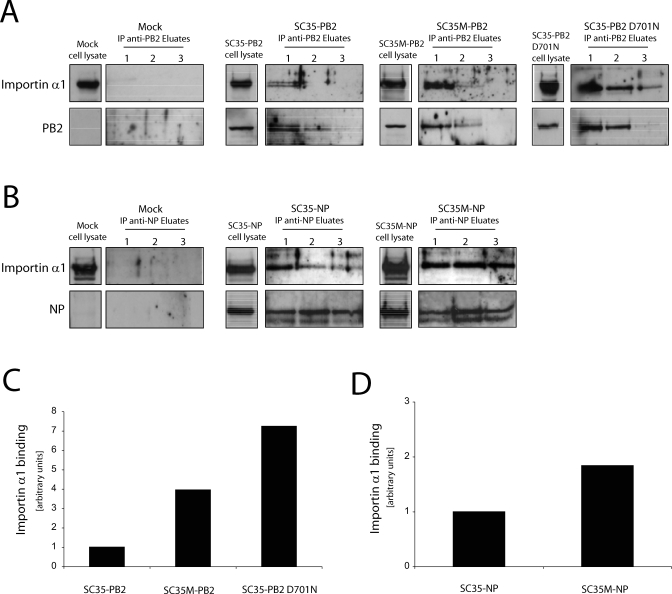
Figure 1. Interaction of PB2 and NP with Importin α1 in Mammalian Cells.
293T cells were transfected with plasmids encoding SC35-PB2 (30 μg), SC35M-PB2 (30 μg), and SC35-PB2 D701N (30 μg) (A) and SC35-NP (10 μg) and SC35M-NP (10 μg) (B) proteins. As a control, non-transfected cells were used (Mock). 48 h after transfection cells were harvested and immunoprecipitated using the respective PB2 or NP antibody. Precipitated eluates (1–3) and non-precipitated whole cell lysates of transfected cells were subjected to SDS gel electrophoresis, and PB2, NP, and importin α1 were detected by Western blot analysis. In (B) two NP bands can be seen of which the minor one most likely represents a cleavage product [39]. The total amount of importin α1 bound in all three eluates to PB2 (C) and NP (D) was quantitated using Image J software (http://rsb.info.nih.gov/ij/). An arbitrary unit is the amount of importin α1 bound to SC35 PB2 and SC35 NP, respectively.

