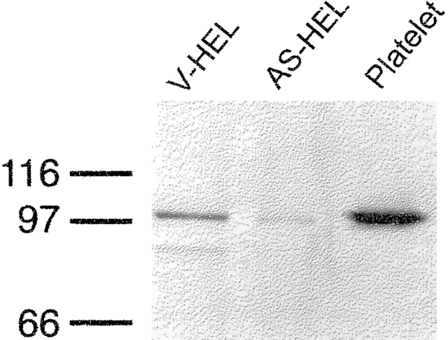
Figure 1.
Western blot demonstrating reduced expression of the GAP1IP4BP in AS-HEL cells. V-HEL and AS-HEL lanes were loaded with 40 μg of the cell lysate from V-HEL and AS-HEL cells and separated by SDS PAGE. A polyclonal anti– GAP1IP4BP antiserum was used as a probe. A platelet plasma membrane fraction served as a positive control for the antibody staining (Platelet).

