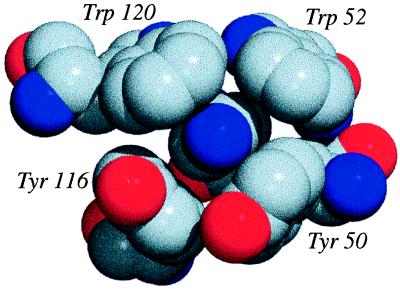
Figure 6.
A cluster of cation-π interactions from the protein glucoamylase (PDB ID code: 1GAI). The NH3+ of the lysine (central blue sphere; Hs not shown) is surrounded by two tryptophans and two tyrosines that contribute −22 kcal/mol Ees. The figure was generated by using povchem (http://grserv.med.jhmi.edu/∼paul/PovChem.html) and povray (http://www.povray.org/). Gray, carbon; red, oxygen; blue, nitrogen.

