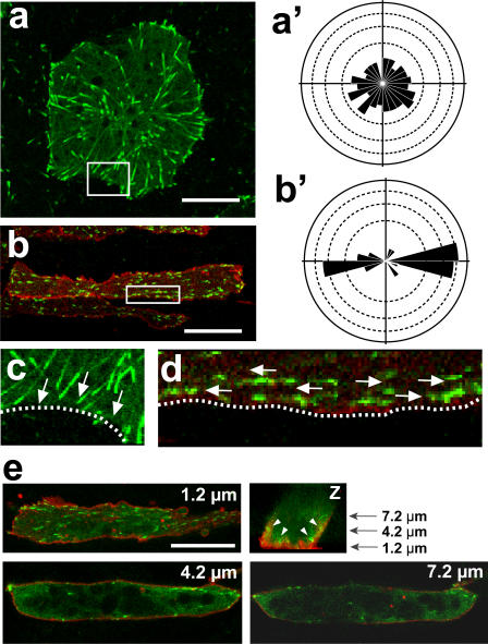
Figure 1. Tracking of EB3-GFP movement shows microtubule growth in Xenopus explants.
(a) In an animal cap cell, EB3-GFP molecules show radially symmetrical movement toward the rim of the cell in the rose diagram (a'). (b) In a chordamesoderm cell, the movement was mostly bidirectional toward both ends of the cell (b'). (c, d) Magnification of the membrane region (white box) of a and b shows that EB3 moved toward the cytoplasmic membrane and disappeared after attaching to it in an animal cap cell (c), while EB3 moved along the membrane in a dorsal mesoderm cell. Arrows show the direction of the EB3 movement, and dotted lines indicate the cytoplasmic membrane. (d). (e) Z section of chordamesoderm cell reveals that most of the EB3 comets are observed in the plane, which is 1.2 µm from glass dish. In the planes, which are 4.2 µm and 7.2 µm from glass dish, the yolk granule were located in the center region of the cell and EB3 comets were located only near the cytoplasmic membrane (white arrow heads). Scale bars, 20 µm. Each scale of the rose diagrams corresponds to 10%.

