
Fig 3.
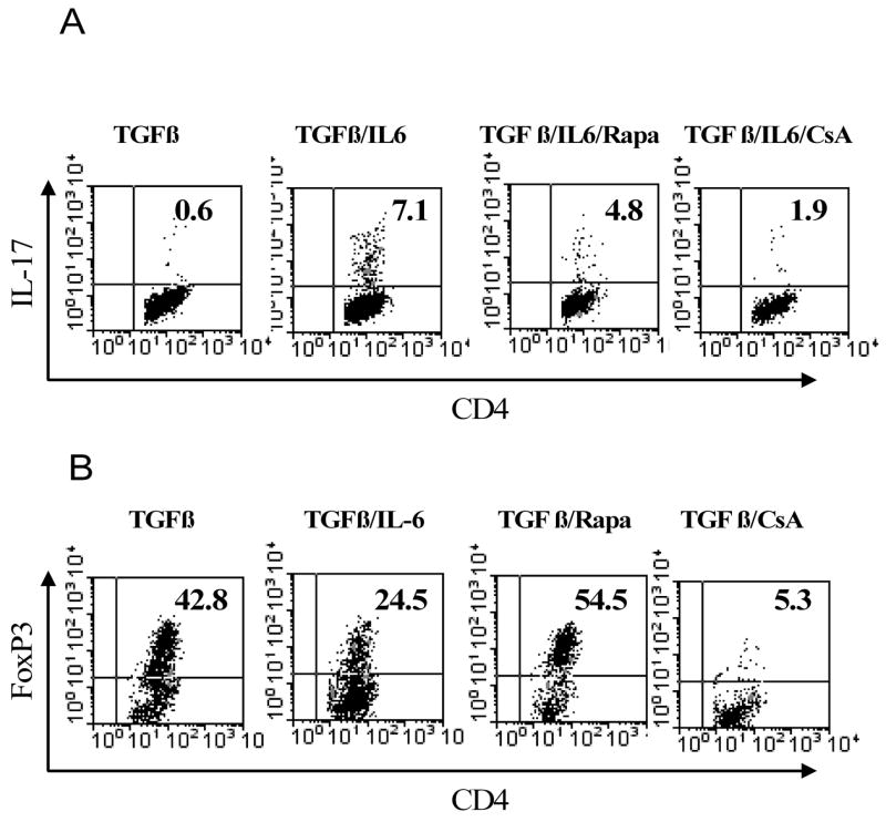
The effects of rapamycin and CsA on generation of FoxP3+ cells and IL-17-producing cells from CD4+CD25− cells. CD4+CD25− cells were isolated from normal C57BL/6 mouse LNs and spleen by flow cytometry. (A) The cells were stimulated with plate-bound anti CD3 Ab and soluble anti CD28 Ab, in the presence of TGFβ, or TGFβ plus IL-6, or TGFβ plus IL-6 plus rapamycin (Rapa, 10 ng/ml), or TGFβ plus IL-6 plus CsA (10 nM). (B) in the parallel experiments, the cells were stimulated with plate-bound anti CD3 Ab and soluble anti CD28 Ab, in the presence of TGFβ, or TGFβ plus IL-6, or TGFβ plus IL-6 plus rapamycin (Rapa, 10 ng/ml), or TGFβ plus IL-6 plus CsA (10 nM). 72 hours later, intracellular expression of FoxP3 or IL-17 was determined by FACS. Analysis was gated on CD4 cells. The data shown are representative of at least 3 separate experiments with similar results.