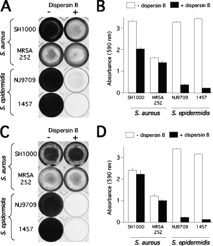
FIG. 1.
Inhibition and detachment of S. aureus and S. epidermidis biofilms by dispersin B in 96-well microtiter plates. (A) The indicated strains were grown for 24 h in unsupplemented TSB medium (−) or TSB supplemented with 20 μg/ml of dispersin B (+). Wells were rinsed and stained with crystal violet. (B) Quantitation of crystal violet staining in panel A. Wells were destained with acetic acid, and the absorbance of the crystal violet solution was measured at 590 nm. Absorbance is proportional to biofilm biomass. Values represent the means for duplicate wells, and error bars indicate range. (C) Biofilms were grown for 24 h in unsupplemented TSB and then rinsed, treated for 1 h with PBS (−) or PBS with dispersin B (+), rinsed, and stained with crystal violet. (D) Quantitation of crystal violet staining in panel C.

