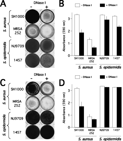
FIG. 4.
Inhibition and detachment of S. aureus and S. epidermidis biofilms by DNase I in 96-well microtiter plates. (A) The indicated strains were grown for 24 h in unsupplemented TSB medium (−) or TSB supplemented with 100 μg/ml of DNase I (+). Wells were rinsed and stained with crystal violet. (B) Quantitation of crystal violet staining in panel A as described in the legend to Fig. 1. (C) Biofilms were grown for 24 h in unsupplemented TSB and then rinsed, treated for 1 h with DNase I buffer alone (−) or DNase I buffer with DNase I (+), rinsed, and stained with crystal violet. (D) Quantitation of crystal violet staining shown in panel C.

