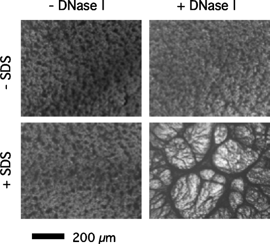
FIG. 5.
Close-up view of 24-h-old S. epidermidis NJ9709 biofilms in 96-well microtiter plates. Top left, untreated biofilm; top right, biofilm treated with 100 μg/ml of DNase I for 1 h; bottom left, biofilm treated with 1% SDS for 5 min; bottom right, biofilm treated with DNase I for 1 h and then SDS for 5 min. Biofilms were stained with crystal violet and then viewed and photographed under an Olympus IMT-2 inverted microscope at magnification ×40.

