FIG. 1.
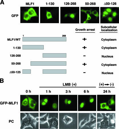
The cytoplasmic localization of MLF1 is disrupted by LMB. (A) Subcellular localization and schematic representation of MLF1 deletion mutants. 293T cells were transfected with the GFP-MLF1/WT and GFP-MLF1 mutant expression vectors and viewed by using fluorescence (GFP) microscopy (upper panels). Results of growth inhibition and subcellular localization are summarized on the right (lower panel). (B) Effects of LMB treatment on the subcellular distribution of MLF1. 293T cells transfected with GFP-MLF1 were treated with 10 ng/ml of LMB and photographed at 1, 3, and 6 h by fluorescence (GFP) microscopy. Cells were also photographed at 24 h after the removal of LMB from the culture. PC, phase contrast.

