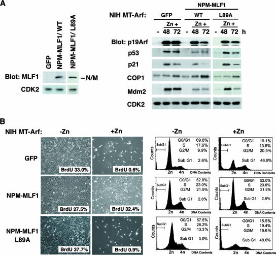
FIG. 6.
NPM-MLF1 fusion protein prevents full induction of p53 by oncogenic cellular stress. The inducible NIH MT-Arf cells were transfected with GFP control, GFP-fused NPM-MLF1, and NPM-MLF1/L89A expression vectors and then selected in puromycin, followed by cell sorting. (A) Expression levels of the NPM-MLF1 (WT and L89A) proteins are shown by immunoblotting with an antibody to MLF1 (left panel). Cells treated with zinc for the indicated times were harvested and analyzed by immunoblotting with antibodies to p19Arf, p53, p21, COP1, Mdm2, and CDK2 (right panel). (B) NIH MT-Arf cells treated with (+Zn) and without (-Zn) zinc for 96 h were photographed (left panel). At 72 h post-HA-Arf induction, cells were subjected to a BrdU incorporation assay (left panel) and a flow cytometric analysis of the DNA content (right panel) for cell cycle analysis. More than 500 BrdU-stained cells were enumerated. Percentages of BrdU uptake are shown at the bottom right of each photograph (left panel).

