FIG. 7.
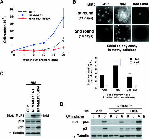
Growth advantage and p53 instability of hematopoietic cells expressing NPM-MLF1. Freshly isolated BM cells were infected with retroviruses expressing GFP alone or GFP-fused NPM-MLF1 or NPM-MLF1/L89A protein. (A) Growth curve for BM cells. Infected BM cells were selected in puromycin and maintained in BM medium. Cells were enumerated and split every 4 days. Data are averages of three independent experiments shown as means ± standard deviations. (B) GFP-positive cells in infected BM cells were isolated by cell sorting and plated in a methylcellulose-based medium. Colonies were viewed after 3 weeks (upper panels, “1st round”) (original magnification, ×16). Viable cells were harvested, counted (lower panel, 1st), and replated in a fresh medium. Colonies were viewed (upper panels), and viable cells were enumerated (lower panel) after 2 weeks (“2nd round”). (C) Infected BM cells, which were selected and maintained in BM medium as shown in panel A, were harvested after 3 weeks postinfection. Expression levels of NPM-MLF1, p53, p21 and γ-tubulin were shown by immunoblotting. (D) Cells, selected and maintained as described in the legend to panel C, were treated with 25 J/m2 of UV irradiation, and p53, p21 and γ-tubulin expression levels were measured by immunoblotting at the indicated times after exposure. Results shown in panels C and D are representative of two independent experiments.

