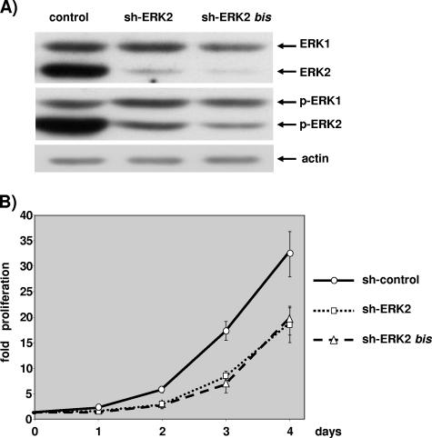
FIG. 3.
ERK2 ablation slows cell proliferation. NIH 3T3 cells were transfected with 3 μg of pBabePuro plasmid and 27 μg of control plasmid, 27 μg pSUPER-ERK2 (sh-ERK2), or 27 μg pSUPER-ERK2bis plasmids (sh-ERK2bis). After selection, cells were plated under conditions of exponential growth. (A) The levels of ERKs and phosphorylated ERKs were evaluated by immunoblotting at 2.5 days postplating as for Fig. 2. The level of actin analyzed by immunoblotting is given for loading normalization. (B) Cells plated on 12-well plates were fixed and nuclei counted as described for Fig. 2. These data are representative of at least four independent experiments. Values are as described for Fig. 2.

