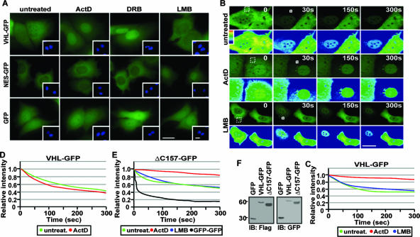
FIG. 1.
Transcription-dependent nuclear export of VHL. (A) ActD alters the steady-state localization of VHL. MCF-7 cells transiently expressing VHL-GFP, NES-GFP, or GFP were either untreated or treated with LMB (10 μM), ActD (8 μM), or DRB (25 μg/ml). Insets are the corresponding Hoechst staining of the cells. Bars, 10 μm. (B and C) Cytoplasmic FLIP reveals that ActD decreases nuclear export of VHL. MCF-7 cells transiently expressing VHL-GFP were treated with ActD (2 μM) or LMB (10 μM) for 1 h. Cells were initially bleached in a large cytoplasmic region (dashed squares) to reduce cytoplasmic signal and then submitted to repetitive bleaching in a small cytoplasmic region (white squares). Cells were imaged between pulses, and the corresponding kinetics for loss of nuclear fluorescence were calculated and plotted on a graph (C) (see Materials and Methods). Bar, 10 μm. (D) Nuclear FLIP analysis was performed on cells treated as for panel B by repetitive bleaching in a small area in the nucleus. The loss of nuclear fluorescence was graphed. (E) ΔC157 exports from the nucleus in a transcription-dependent manner. ΔC157-GFP-transiently expressing cells were treated and analyzed as described for panel B. (F) Western blot analysis using anti-Flag and anti-GFP antibodies verified that the Flag- and GFP-tagged VHL, ΔC157, and GFP fusion proteins used for photobleaching experiments were not fragmented.

