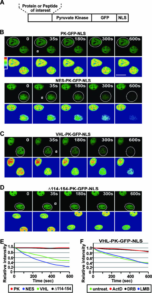
FIG. 2.
A transcription-dependent nuclear export sequence is encoded within the exon-2-encoded β-domain of VHL. (A) Schematic diagram of the PK-GFP-NLS fusion protein used for the live cell FLIP nuclear export assay, describing the region where protein or peptide sequences were fused. (B to E) MCF-7 cells transiently expressing the indicated constructs were submitted to cytoplasmic FLIP analysis, in which a small cytoplasmic region (white squares) within specific cells (a dashed circle outlines the cell nucleus) was repeatedly bleached. Kinetics for the loss of nuclear fluorescence from images obtained in panels B, C, and D were calculated and plotted on a graph (E). PK refers to the PK-GFP-NLS reporter construct, and NES, VHL, and Δ114-254 indicate the sequences fused to PK-GFP-NLS. Bar, 10 μm. (F) Cells were transiently transfected with VHL-PK-GFP-NLS and were either treated with ActD (2 μM), DRB (25 μg/ml), or LMB (10 μM) for 1 h or left untreated. Cytoplasmic FLIP was performed as described above to verify nuclear export activity.

