FIG. 8.
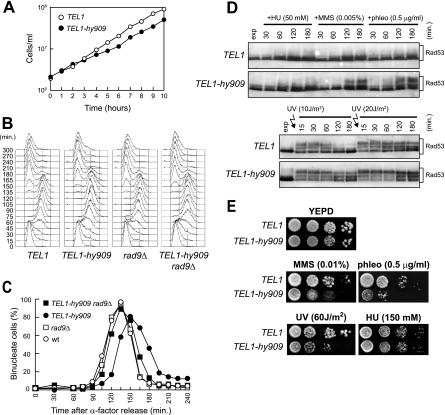
Checkpoint activation in TEL1-hy909 MEC1 cells during an unperturbed cell cycle or in response to mild genotoxic treatments. (A) Growth rate of asynchronous wild-type (W303; TEL1) and TEL1-hy909 cell cultures growing exponentially at 25°C. (B and C) Exponentially growing cell cultures of wild-type (W303; TEL1), TEL1-hy909, rad9Δ (DMP1911/1C), and TEL1-hy909 rad9Δ (DMP4711/3C) strains, all expressing wild-type MEC1, were arrested in G1 with α-factor (0) and released from the pheromone block in YEPD at 21°C. When 95% of cells had budded after release, 3 μg/ml α-factor was added back to all cultures. Samples were collected at the indicated times after α-factor release to analyze the DNA content by a fluorescence-activated cell sorter (B) and to determine the kinetics of nuclear division after propidium iodide staining (C). wt, wild type. (D) Cell cultures of exponentially growing (exp) wild-type (W303; TEL1) and TEL1-hy909 strains were UV irradiated (10 or 20 J/m2) or were resuspended in YEPD containing 50 mM HU (+HU), 0.005% MMS (+MMS), or 0.5 μg/ml phleomycin (+phleo). Protein extracts prepared from cell samples collected at the indicated times after genotoxic treatment were subjected to Western blot analysis with anti-Rad53 antibodies. (E) Serial dilution of wild type (W303; TEL1) and TEL1-hy909 cell cultures, exponentially growing in YEPD, were spotted on plates with or without phleomycin, MMS, and HU at the indicated concentrations. One YEPD plate was exposed to the indicated UV dose.

