FIG. 4.
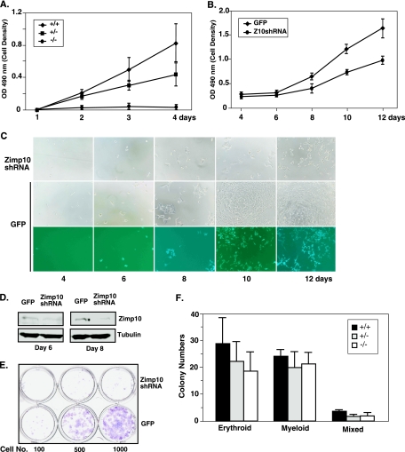
Effects of Zimp10 loss on cell viability and hematopoietic potential. (A) MTS cell proliferation assay using E9.5 MEFs isolated from zimp10 heterozygous intercrosses (+/+, n = 4; +/−, n = 4; −/−, n = 3). (B to E) HEK293 cells were infected with Zimp10 shRNA (Z10shRNA) lentivirus or GFP control virus and replated for the assessment of cell viability using the MTS cell proliferation assay (B), bright-field and fluorescence microscopy (C), or clonogenic assay (E). (D) Western blot analysis of the knockdown effect in cells after 6 or 8 days of infection with Zimp10 shRNA (top panel). The same blots were probed with an antitubulin antibody as a control. (F) E9.5 yolk sacs were isolated from heterozygous intercrosses, disaggregated, and plated for hematopoietic colony-forming ability on methocellulose semisolid medium plus hematopoietic growth factors (5 × 10⁴ cells/well). Erythroid colonies were counted on day 4, and myeloid colonies were counted on day 11. Erythroid-myeloid mixed colonies were counted accordingly. Bars represent averages and standard deviations of results from multiple yolk sacs isolated from the same litter (Zimp10+/+, n = 6; Zimp10+/−, n = 7; Zimp10−/−, n = 6). OD 490 nm, optical density at 490 nm.

