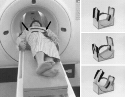
Fig. 1.
Acquisition of 3D-MRI. Our original device (arrow) was used to rotate the trunk of the subjects as reproducibly as possible. 3D MRIs were obtained in nine positions with 15° increments of trunk rotation (0°, 15°, 30°, 45°, and maximum). Maximum trunk rotation was defined as the position in which the trunks were rotated as much as possible without the bilateral shoulders pulled away from the MRI table

