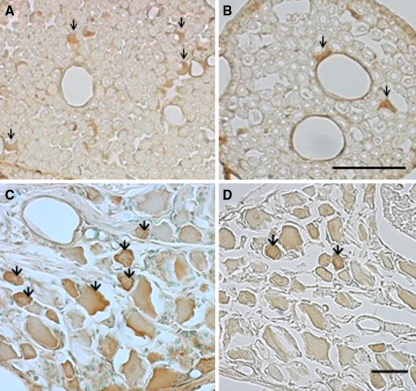
Fig. 3.
Photomicrographs demonstrating VEGF-immunoreactive cells in the cauda equine (a, b) and DRG (c, d) in compression group (a, c) and sham-operated group (b, d) at day 28 after surgery. Schwann-like cells in the cauda equina and DRG neurons exhibited immunoreactivity for VEGF (arrows). Scale bar 50 μm

