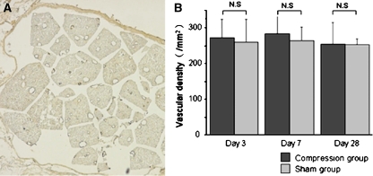
Fig. 5.
a Photomicrographs demonstrating Factor VIII-immunoreactive blood vessels in the cauda equina of sham-operated rat at day 3 after surgery. Blood vessels are visible after immunostaining for factor VIII. Scale bar 200 μm. b Histogram presenting vascular density in the cauda equina. There were no significant differences between the compression and sham-operated groups at any time point. Results are the mean ± standard deviation of vascular density

