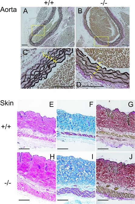
FIG. 2.
Histological analysis of ascending aortae and skin from Fbln2+/+ and Fbln2−/− mice. (A to D) Paraffin-embedded aortic sections were stained with Verhoeff's solution, in which elastin appears dark brown or black. Images were taken at two different magnifications. Arrows indicate elastin laminae. Magnification bar = 100 μm. (E to J) Skin sections were subjected to hematoxylin-eosin (E and H), Masson's trichrome collagen (F and I), and Verhoeff's van Gieson elastin (G and J) stains. Bars = 100 μm.

