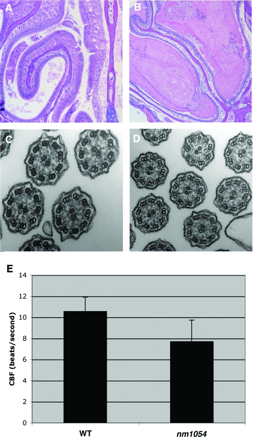
FIG. 3.
Respiratory abnormalities in B6129F1 nm1054 mice. (A and B) Coronal sections of 8-week-old wild-type (A) and nm1054 (B) maxillary sinus and turbinate samples. Note the accumulation of mucus in the nm1054 sinus (B). Sections were stained with hematoxylin and eosin. (C and D) Transmission electron microscopy of wild-type (C) and nm1054 (D) tracheal epithelial cilia, demonstrating a normal axonemal ultrastructure. (E) Ciliary beat frequency (CBF) of wild-type and nm1054 tracheal epithelial cilia, showing a decreased rate of beating in the mutant (n = 6 in each group; P < 0.01).

