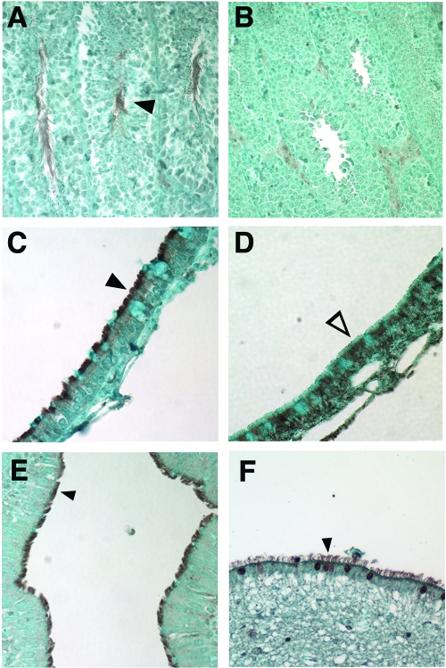
FIG. 7.
Immunohistochemical analysis of mouse and human Pcdp1 protein expression. (A and B) Expression of Pcdp1 in B6129F1 wild-type (A) and nm1054 (B) testes. (C and D) Expression of Pcdp1 in B6129F1 wild-type (C) and nm1054 (D) sinus epithelial cells. (E and F) Expression of Pcdp1 in human ciliated bronchial epithelial cells (E) and brain ependymal cells (F). Closed arrowheads indicate presence of mouse Pcdp1 in wild-type flagella (A) and cilia (C) and human PCDP1 in bronchial epithelial (E) and ependymal (F) cilia. The open arrowhead indicates an absence of Pcdp1 in nm1054 sinus epithelial cilia (D).

