FIG. 5.
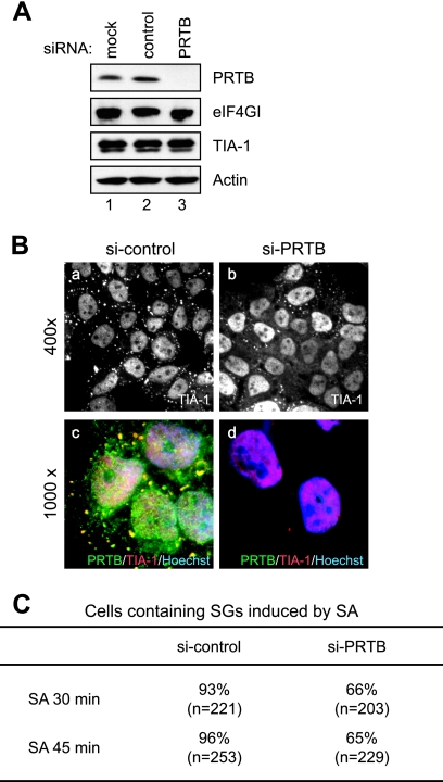
Effect of PRTB knockdown on SG formation. (A) Level of PRTB protein in cells treated with an siRNA against PRTB mRNA. Levels of PRTB, eIF4GI, TIA-1, and actin proteins were analyzed by Western blotting using antibodies against PRTB, eIF4GI, TIA-1, and actin, respectively. HeLa cells were used in the Western blotting before treatment (lane 1) and in treatments with control siRNA (lane 2) and synthetic siRNA against PRTB (lane 3) for 48 h. (B) Effect of siRNA against PRTB on SG formation. HeLa cells were treated with control siRNA (si-control) (images a and c) and siRNA against PRTB (si-PRTB) (images b and d) for 48 h and then were treated with 400 μM of SA for 30 min. SG formation was monitored by immunocytochemistry using anti-TIA-1 antibody (images a and b). Donkey antibodies against rabbit immunoglobulin G conjugated with FITC and against goat immunoglobulin G conjugated with rhodamine were used as secondary antibodies. Merged images of PRTB (green), TIA-1 (red), and Hoechst 33258-stained nuclei (blue) are shown in images c and d. Microscopic magnifications of the pictures are ×400 (images a and b) and ×1,000 (images c and d). (C) Proportions of cells containing SGs. After treatment with SA for 30 and 45 min, HeLa cells were prepared as described for panel B. The numbers of cells containing more than four SGs were counted under a fluorescence microscope. The total numbers of the cells observed are listed in parentheses, and the proportions of SG-containing cells among the observed cells are listed as percentages.

