FIG. 2.
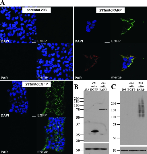
Constitutive presence of PAR in mitochondria of stably transfected 293 cells. (A) Images from confocal scanning of 293 cells stably transfected with mitoPARP-encoding vector (293mitoPARP panels) after indirect immunocytochemistry using 10H antibody to detect PAR. Mitochondrial PAR accumulation colocalizing with the intrinsic EGFP fluorescence of the synthetic PARP construct was detectable in all cells. 293 cells stably transfected with a construct lacking the PARP portion of the construct (293mitoEGFP panels) were EGFP positive but PAR negative, whereas in parental 293 cells (parental 293 panels) neither green fluorescence nor immunoreactivity for PAR was detected. Bar, 20 μm. (B) Forty micrograms of total protein from the indicated cell lines was separated by 10% SDS-PAGE. Western blotting was performed using myc antibody. (C) Forty micrograms of total protein of the indicated cell lines was separated by 7% SDS-PAGE. PAR was revealed by Western blotting using the 10H antibody. β-Tubulin was used as a loading control in panels B and C.

