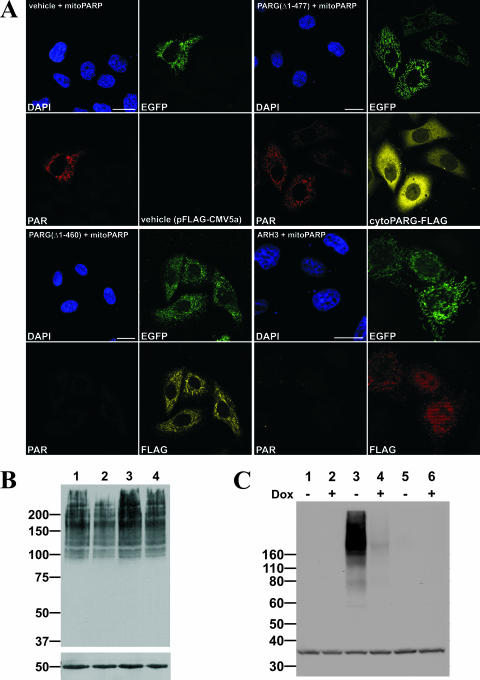
FIG. 6.
PARG(Δ1-460) and ARH3 degrade PAR in mitochondria. (A) Images from confocal laser scanning of HeLaS3 cells transiently cotransfected with vector encoding mitoPARP and either PARG(Δ1-460) harboring a predicted mitochondrial targeting sequence (panels in lower left quadrant) or ARH3 (panels in lower right quadrant). Control cells were cotransfected with mitoPARP-encoding vector and empty plasmid (vehicle; panels in upper left quadrant) and a vector encoding PARG(Δ1-477), which lacks the predicted targeting sequence (panels in upper right quadrant). Bar, 20 μm. (B) Stably transfected 293mitoPARP cells were either transfected with vectors encoding PARG(Δ1-460) (lane 2), PARG(Δ1-477) (lane 3), and empty plasmid (lane 4) or not transfected (lane 1) and subjected to Western blot analysis using 10H antibody. Fifty micrograms of total protein was separated by 7% SDS-PAGE; β-tubulin was used as loading control. (C) Doxycycline-induced (+) and noninduced (−) Flp-In T-REx 293 cells expressing ARH3 under control of a tetracycline-inducible promoter were transfected with the vector encoding mitoPARP (lane 3 and 4). Control cells were transfected with empty vector (lane 5 and 6) or left untransfected (lane 1 and 2). For Western blot analysis of PAR, 50 μg was separated by 4 to 12% SDS-PAGE. As a loading control eukaryotic translation initiation factor 2α was used.

