FIG. 3.
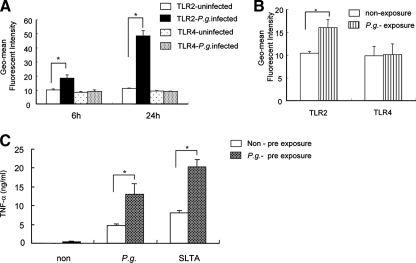
P. gingivalis up-regulates TLR2 surface expression on murine macrophages. (A) Peritoneal macrophages from WT mice were cultured with P. gingivalis (MOI = 10) or with medium alone (uninfected), and the temporal expression levels of TLR2 and TLR4 were determined by FACS analysis at 6 and 24 h. Data are expressed as the geometric (Geo)-mean of fluorescence intensities and standard deviations (SDs) of three independent experiments. *, P < 0.01 by unpaired t test. P.g., P. gingivalis. (B) Peritoneal macrophages from WT mice were cultured with P. gingivalis (MOI = 10) or with medium alone for 1 h, and then nonadherent bacteria were removed by washing out cells. The cells were then incubated with medium alone for 5 h, and the temporal expression levels of TLR2 and TLR4 were determined by FACS analysis. Data are expressed as the geometric mean fluorescence intensities and SDs of three independent experiments. *, P < 0.01 by unpaired t test. (C) Non-preexposed (Non) peritoneal macrophages or macrophages preexposed to P. gingivalis (MOI = 10) for 1 h were washed and incubated with medium alone for 5 h. Following a change of the medium, cells were incubated with medium, with P. gingivalis (MOI = 10), or with SLTA (2 μg/ml) for 24 h, and TNF-α production in the CS fluids was assessed by ELISA. Data are shown as the means and SDs of two independent experiments, each performed in triplicate. *, P < 0.01 by unpaired t test.

