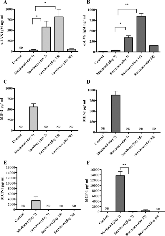
FIG. 2.
Antibody and chemokine responses in lungs and spleens of mice infected with LVS. Following intranasal infection with LVS, mice (three to six animals per group) were humanely sacrificed at a terminal disease stage or at different time points after recovering from symptoms. The results are shown in light gray for spleens and dark gray for lungs. Organs from mice injected with PBS were used as negative controls. Data are presented as the quantity (in ng or pg) of antibody or chemokine per milliliter at different stages of disease, and the error bars represent standard deviations. Differences between moribund mice and survivors were either significant (*, P < 0.05) or highly significant (**, P < 0.01). ND, not detectable.

