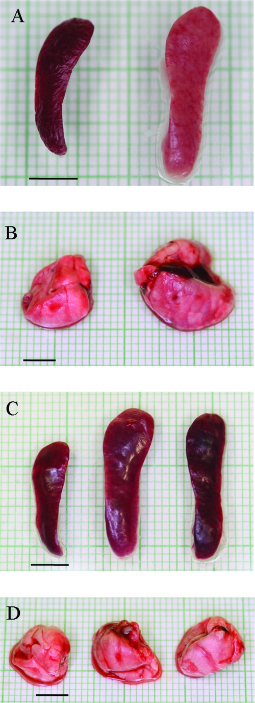
FIG. 4.
Changes in lung and spleen gross anatomy after infection with LVS. (A) Spleens collected from representative moribund mice appeared enlarged and pale (right) compared to those of uninfected controls (left). (B) Lungs from representative moribund mice did not collapse and showed some color variation (right) compared to those from uninfected controls (left). (C) Splenomegaly in representative survivors at early (day 7; center) and late (day 50; right) time points in comparison to normal spleens from uninfected control mice (left). (D) Lung tissue from survivors at both early (day 7; center) and late (day 50; right) times appeared normal in size and color, similar to those of uninfected controls (left). Bars = 5 mm.

