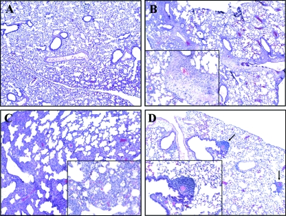
FIG. 7.
Hematoxylin and eosin staining of formalin-fixed pulmonary tissue. (A) No alterations were observed in the lung tissue of uninfected control mice. (B) Seven days after infection, moribund mice presented with pneumonia characterized by diffuse edema, neutrophil infiltration, and fibrin deposits. The inset shows a large area of diffuse edema. (C) Survivors sacrificed at day 7 postinfection presented with severe cellular infiltration but no fibrin deposits or edema. Neutrophil infiltration is evident in the inset. (D) Survivors sacrificed at day 50 no longer showed pathological changes in the lung, and lymphoid aggregates were evidenced, as shown by the arrows. The sections were photographed with a 10× objective, with a 50× objective for the insets.

