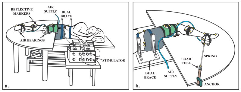
Fig. 1.
Experimental Setup. (a) The lower extremity was supported on air bearings that allowed near frictionless motion in the sagittal plane. Pelvis motion was restricted by a dual brace, padded restraint. An electrical stimulator delivered a pulse train to the rectus femoris, the vastus medialis or both muscles simultaneously. Reflective markers were used to measure the induced lower extremity kinematics via an 8-camera motion capture system. (b) Compliant springs, attached to fixed load cells, were used to hold the limb in a desired posture prior to stimulation.

