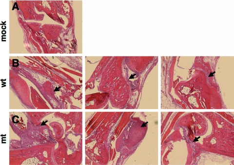
FIG. 5.
Histological examination of joints from C3H/HeN mice infected intradermally with 103 spirochetes per mouse. The tibiotarsal joints were collected 62 days postinfection, processed as described in Materials and Methods, and stained with hematoxylin and eosin. The joint-tissue samples are from mice inoculated with phosphate-buffered saline (mock), the wt (ML23/pBBE22; n = 3), or the mt (MM4/pBBE22; n = 3). Note the presence of increased levels of infiltration in joint tissue of mice (samples 1 and 2) inoculated with the mt compared to that of mice inoculated with the wt, as indicated by arrows.

