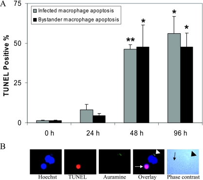
FIG. 1.
M. tuberculosis strain H37Ra induces bystander apoptosis in uninfected human macrophages. (A) Macrophages were infected with M. tuberculosis strain H37Ra (47.95% ± 12.05% cells were infected with an MOI of 1 to 5 bacilli per cell) for 4 h in two-well LabTek chambers. Apoptosis was determined for both infected and uninfected bystander cells after 24, 48, and 96 h by the in situ TUNEL TMR technique. Significant DNA fragmentation occurred in H37Ra-infected macrophages at 48 h (P < 0.005) and 96 h (P < 0.05) postinfection compared to the control. The uninfected bystander apoptosis was significantly greater than the apoptosis of uninfected control cells at 48 h (P < 0.05) and 96 h (P < 0.05) postinfection. The data are expressed as percentages of TUNEL-positive cells (means and standard errors of the means) of triplicate samples, and at least 100 cells per slide were counted at a magnification of ×100. Statistical significance was determined using the Student t test (one asterisk, P < 0.05; two asterisks, P < 0.005). (B) Fluorescent microscopy images showing a TUNEL-positive, apoptotic uninfected bystander macrophage (white arrow) in close proximity to a nonapoptotic infected cell whose nucleus was stained using Hoechst stain. The white arrowhead indicates intracellular auramine-stained (green) bacilli. The phase-contrast light micrograph shows an infected cell (black arrowhead) and uninfected bystander cell (black arrow) in contact.

