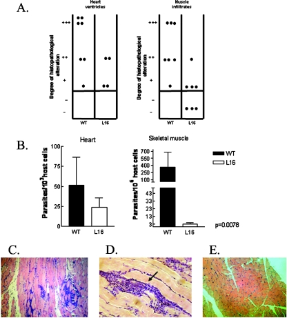
FIG. 6.
Inflammatory infiltrates and tissue parasitism in the heart and skeletal muscles of mice chronically infected with either the L16 clone or the WT CL Brenner strain. (A) Histopathological analysis was plotted as dispersion diagrams indicating the intensities of inflammatory infiltrates graded as absent (−), slight (+), moderate (++), and severe (+++). Each dot represents a mouse. Mice inoculated with 103 CRF were autopsied at 7 months postinfection. The L16 group showed a significantly low inflammatory response in muscle tissue (P < 0.05) and a low degree of inflammation in heart tissue compared to the levels of the WT group. (B) Parasite burden in skeletal and cardiac muscles at the chronic phase of infection. Data are expressed as the number of parasites for 106 or 103 host cells. (C and D) Microphotograph of muscle from mice infected with WT strain; note the amastigote nest inside the inflammatory infiltrates (arrow) (magnification, ×25). Values are given as means; error bars indicate standard errors of the means. (E) Muscle of L16-infected mouse, showing the absence of dense inflammatory infiltrates relative to the WT group.

