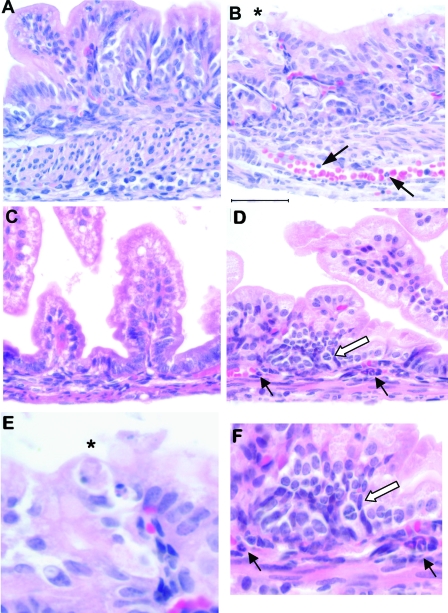
FIG. 5.
Inflammatory changes in the GI tract of critically ill SP-A null mice exposed to corn dust bedding. Twenty-four-hour-old SP-A null mice born and reared in control or corn dust bedding were examined. Representative histological sections from stomachs (A, B, and E) and small intestines (C, D, and F) are shown. SP-A null mice born in corn dust bedding (B) frequently exhibited sloughing gastric epithelial cells with condensed nuclei (asterisk) compared to SP-A null mice born in control bedding (A). On average, we observed 0.80 ± 0.26 sloughing gastric cell/high-power field in the SP-A null mice, which is significantly more (P < 0.05) than we observed in the SP-A null mice exposed to control bedding (0.25 ± 0.13 cell/high-power field). An example of this is shown in panel E. Neutrophils were also observed within and marginating along the gastric blood vessels of critically ill SP-A null pups in corn dust bedding (arrows in panel B). Also, SP-A null mice reared in corn dust bedding frequently exhibited inflammatory changes in the proximal small bowel (D) compared to SP-A null mice reared in control bedding (C). Specifically, neutrophils were frequently observed within the blood vessels at the base of the villi and also outside blood vessel walls (black arrows in panel D). Additionally, collections of mononuclear cells were also observed only in the ill SP-A null newborns exposed to corn dust bedding (open arrow in panel D). These changes in the proximal small bowel are shown at a higher magnification in panel F. These inflammatory findings were not observed in any of the age-matched SP-A null mice born in control bedding. The images are representative of four to six mice per condition. Bar = 50 μm.

