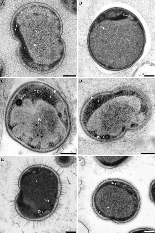
FIG. 2.
Transmission electron micrographs of high-pressure frozen, freeze-substituted, and Epon-embedded thin sections of four anammox genera. All cells are divided into three separate compartments by individual membranes: the paryphoplasm (p), riboplasm (r), and anammoxosome (a) compartments. (A) Dividing “Candidatus Kuenenia stuttgartiensis” cell. (B) “Candidatus Anammoxoglobus propionicus” cell. (C and D) “Candidatus Brocadia fulgida” cells showing riboplasmic particles (pa). (D) Dividing cell. (E and F) Dividing “Candidatus Scalindua spp.” cells with (E) and without (F) pilus-like appendages. Micrographs further show glycogen (g) and putative intra-anammoxosome iron particles (Fe). Scale bars, 200 nm.

