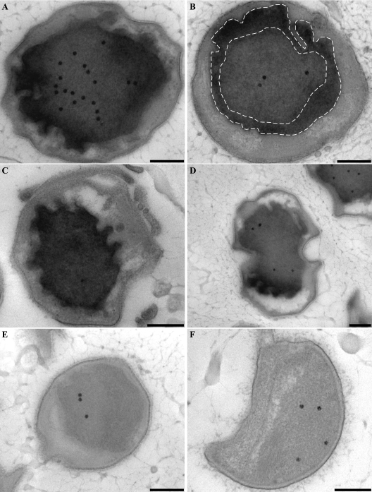
FIG. 5.
Transmission electron micrographs of chemically fixed and Epon-embedded thin sections of “Candidatus Kuenenia stuttgartiensis” cells showing cytochrome peroxidase staining. This figure is best viewed on screen to avoid change of contrast by printer settings. (A to D) Cytochrome peroxidase staining is observed solely in the anammoxosome compartment. Intense staining occurs within close proximity to the anammoxosome membrane, as outlined by the dashed lines (B) and in places where the membrane is curved (D). (E and F) Negative controls preincubated with KCN and incubated with DAB and H2O2 in the presence of KCN. All sections were poststained for 1 min with Reynolds lead citrate. Scale bars, 200 nm.

