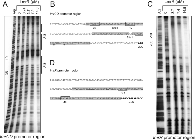
FIG. 3.
DNase I protection of the lmrCD and lmrR promoter regions by LmrR, showing site specificity of binding of LmrR to the lmrCD (A and B) and lmrR (B and D) promoter regions. The DNase I-digested promoter fragments (A and C) are flanked by the Maxam-Gilbert ladder on the left (AG). Poly(dI-dC) was present to suppress unspecific binding. Nucleotide sequences of the lmrC (B) and lmrR (D) promoter regions show the LmrR-protected regions (shaded), the putative −35 and −10 regions (boxed), the inverted repeats (arrows), and the structural genes (bold).

